Home > Conditions > Vascular & Pigmented Lesions
Vascular & Pigmented Lesions Treatment Singapore
Vascular & Pigmented Lesions Treatment Singapore
Vascular and pigmented lesions encompass a broad spectrum of skin conditions characterised by abnormal blood vessels or pigment distribution. These lesions can be present from birth (congenital) or develop over time (acquired). They range from entirely benign cosmetic concerns to lesions requiring careful monitoring or active treatment. Accurate diagnosis is essential, as the management approach varies significantly depending on the specific type of lesion.
At Skincodes, Dr Ang Sue-May provides expert assessment of vascular and pigmented lesions, utilising dermoscopy and clinical expertise to ensure appropriate diagnosis and individualised treatment planning.
Types of Vascular Lesions
Congenital Vascular Lesions
Infantile haemangiomas – Benign vascular tumours appearing shortly after birth, growing rapidly then slowly regressing
Port wine stains – Capillary malformations present at birth, persisting and often darkening over time
Salmon patches – Flat pink patches common in infants, typically fading spontaneously
Acquired Vascular Lesions
Spider angiomas – Central red spot with radiating vessels, common in pregnancy and liver disease
Cherry angiomas – Small, bright red dome-shaped papules, very common with ageing
Telangiectasia – Visible dilated blood vessels, associated with sun damage, rosacea, or genetics
Venous lakes – Dark blue to purple papules, commonly on lips and ears
Pyogenic granulomas – Rapidly growing, bleeding vascular lesions often following minor trauma
Types of Pigmented Lesions
Melanocytic Lesions
Melanocytic naevi (moles) – Benign collections of melanocytes; junctional, compound, or dermal types
Café-au-lait macules – Flat, evenly pigmented light-brown patches; single lesions are common and benign
Congenital melanocytic naevi – Moles present at birth, varying from small to giant
Blue naevi – Blue-black papules resulting from deep dermal melanin
Lentigines – Flat brown spots including solar lentigines (sun spots) and simple lentigines
Non-Melanocytic Pigmented Lesions
Seborrhoeic keratoses – Common ‘age spots’ or ‘barnacles’ with waxy, stuck-on appearance
Dermatofibromas – Firm nodules, often with pigment change and dimple sign
Post-inflammatory hyperpigmentation – Dark marks following skin inflammation or injury
Drug-induced pigmentation – Medication-related colour changes
Signs & Symptoms Requiring Assessment
Whilst many vascular and pigmented lesions are benign, certain features warrant specialist evaluation:
- Changing lesions – Any alteration in size, shape, or colour
- Irregular borders – Notched, blurred, or poorly defined edges
- Multiple colours – Variegation within a single lesion
- Bleeding or ulceration – Without clear trauma
- Rapid growth – Particularly in adulthood
- New onset – Lesions appearing in adulthood
- Symptomatic lesions – Itching, pain, or tenderness
- Cosmetic concern – Lesions affecting appearance or confidence

Diagnosis at Skincodes
Clinical Examination
Dr Ang conducts thorough visual inspection, assessing lesion characteristics including:
- Morphology – Size, shape, surface characteristics
- Colour – Uniform or varied pigmentation
- Texture – Smooth, rough, raised, or flat
- Distribution pattern – Localised or widespread
- Associated features – Surrounding skin changes, satellite lesions
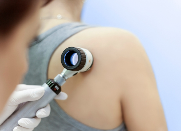
Dermoscopy
This non-invasive technique uses magnification and polarised light to visualise subsurface structures:
- Vascular patterns – Identifying vessel types and arrangements
- Pigment patterns – Assessing pigment network, dots, globules, and structures
- Improved diagnostic accuracy – Distinguishing benign from suspicious features
- Documentation – Photography for monitoring changes over time
When Biopsy is Necessary
If features raise concern, a skin biopsy provides definitive diagnosis:
- Punch biopsy – Full-thickness sample for histological examination
- Shave biopsy – Superficial sampling for raised lesions
- Excision biopsy – Complete removal of suspicious lesions
Treatment at Skincodes
Laser Therapy for Vascular Lesions
Modern laser technology offers effective, targeted treatment:
Yellow laser (585nm) – effective for facial telangiectasia, diffuse redness, and multiple superficial vascular lesions.
Laser Therapy for Pigmented Lesions
Pico laser – Ultra-short pulses for lentigines and selected pigmented lesions
Q-switched laser – For deeper pigmentation
CO₂ laser – Ablative removal of raised seborrhoeic keratoses
Yellow Light Laser – For vascular components of mixed lesions
Surgical Options
Excision – Complete removal of suspicious or cosmetically bothersome lesions
Shave excision – For raised benign lesions
Curettage and cautery – For selected superficial lesions
Conservative Management
Not all lesions require active treatment. Dr Ang provides:
- Reassurance – When lesions are clearly benign
- Monitoring plans – For lesions best observed over time
- Patient education – Warning signs to watch for
- Photographic documentation – For objective comparison at follow-up
What to Expect
Treatment planning depends on lesion type, location, and patient factors:
- Consultation – Comprehensive assessment and diagnosis
- Treatment selection – Matched to lesion characteristics and patient goals
- Multiple sessions – Often required for optimal results
- Healing time – Varies by treatment modality
- Realistic expectations – Some lesions respond better than others; recurrence is possible

Why Choose Specialist Surgical Care
Dermatological surgery performed by a specialist offers distinct advantages:
- Accurate pre-surgical diagnosis with dermoscopy. Lesions are assessed carefully as appearances can be
misleading without specialist evaluation - Appropriate margin assessment for cancer excisions based on lesion type and guidelines
- Histological analysis of all removed tissue to confirm diagnosis and clear margins
- Minimally invasive techniques selected to optimise cosmetic outcomes
- Structured follow-up and surveillance for ongoing skin health