Uneven skin tone is among the most common reasons people see a dermatologist in Singapore, where National Environment Agency data put the daily UV index at 6 to 9 for most of the year. The types of facial pigmentation can look identical yet behave very differently beneath the surface. This blog walks you through how a dermatologist tells them apart and why that distinction decides how each mark is treated.
What are the main types of facial pigmentation?
Facial pigmentation is excess or uneven melanin in the skin, and on Singaporean faces it falls into four common patterns: melasma, solar lentigines (sunspots), post-inflammatory hyperpigmentation, and freckles. Each comes from a different trigger, sits at a different depth, and responds to a different treatment. That is why one “brown spot” label hides four separate problems.
Melanin is the pigment made by melanocytes in the basal layer of the epidermis, and tyrosinase is the enzyme that switches its production on. When that process runs too hard in one area, pigment clusters and becomes visible.
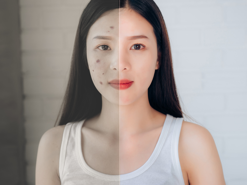
Melasma shows up as symmetrical brown patches across the cheeks, forehead, and upper lip, driven by a mix of hormones and sunlight. Solar lentigines are discrete, well-edged spots from years of accumulated UV. Post-inflammatory hyperpigmentation is the flat brown mark left behind after acne, eczema, or a procedure settles. Freckles are small, light tan spots that darken in sun and fade in winter, often inherited and appearing early in life. The first job in clinic is naming which one you actually have, because the treatment paths diverge from there.
What is the difference between melasma and sunspots?
Melasma is hormonally influenced and symmetrical; sunspots are purely sun-driven and scattered. That single distinction changes the whole treatment plan. Melasma sits on both cheeks in a mirror-image pattern and flares with pregnancy, oral contraceptives, and heat. Solar lentigines appear as separate, sharply bordered spots on the highest sun-exposure zones: the cheekbones, the bridge of the nose, the forehead.
The demographics differ too. Melasma overwhelmingly affects women; a multicentre Brazilian study put the female share at 97.5%, and it concentrates in Fitzpatrick skin types III to V, the range most Singaporean skin falls into. Solar lentigines track age and sun history instead, becoming near-universal in heavily sun-exposed older skin.
Singapore’s climate sharpens both. The National Environment Agency records a daily maximum UV index that frequently reaches “very high” between 11am and 3pm, yet a National Skin Centre survey found only 23.9% of adults here use sunscreen. Constant UV feeds new pigment and re-darkens treated pigment, which is why protecting against cumulative sun damage matters as much as any laser. The practical difference: sunspots can often be cleared and kept away, while the way melasma behaves means it is managed and prone to return rather than “removed” once.

What does epidermal versus dermal pigmentation mean, and why does it matter?
Depth decides how well pigmentation responds, so dermatologists classify it as epidermal, dermal, or mixed before choosing a treatment. Epidermal pigment sits in the upper skin layers, the basal and suprabasal epidermis, where it is more accessible and tends to lighten faster. Dermal pigment lies deeper in the dermis, reads as a grey-blue tone, and clears slowly and incompletely. Mixed pigmentation carries both.
The reason this matters is prognosis. Epidermal melasma can respond well to topical tyrosinase inhibitors and conservative light-based work. Dermal pigment resists creams and needs a different, more cautious strategy. Promising the same timeline for both sets a patient up for disappointment.
This is also where the depth question feeds straight into safety. Deep pigment in darker phototypes is exactly the situation where over-aggressive treatment backfires, because the same energy that breaks up surface pigment can inflame the dermis and lay down fresh pigment. In practice, the depth assessment is not academic. It is the difference between a plan that works and one that makes the face look worse for months.
How do dermatologists diagnose the type of pigmentation?
Diagnosis runs in a fixed order: clinical history, then a Wood’s lamp examination, then dermoscopy, and a biopsy only when something looks off. Each step answers a specific question that the naked eye cannot.
The Wood’s lamp is a near-century-old ultraviolet tool that localises melanin by depth. Epidermal pigment accentuates and looks darker and sharper under the lamp; dermal pigment does not change. That single observation, first formally described by Gilchrest and Fitzpatrick in 1977, sorts a patch into epidermal or dermal in seconds. Dermoscopy then magnifies the pigment network to confirm the pattern and to flag anything suspicious; in one comparative study, Wood’s lamp and dermoscopy findings agreed at a kappa of 0.81, which is substantial but not total, so clinicians use them together rather than alone.
The honest part is that pigmentation is hard to read, even for specialists. When the Asian Acne Board asked seven dermatologists to grade the same clinical photographs, the panel reported “marked variability between raters” in scoring post-inflammatory hyperpigmentation. That variability is the argument for structured diagnosis over a glance. A consistent sequence, not a single tool, is what makes the call reliable.
Why can the wrong treatment make pigmentation worse?
The wrong laser, or the right laser at the wrong setting, can darken melasma and trigger fresh pigment, especially in Fitzpatrick III to V skin. This is the single most important reason diagnosis comes before any device is switched on. Melasma in particular is heat- and inflammation-sensitive: treatments that suit a stable sunspot can destabilise it.
The mechanism is straightforward. Melanocytes in more pigmented skin are more reactive. Hit them with excess energy and they respond by producing more melanin, not less, which is how a patient ends up with deeper, wider discolouration than they started with. Worth noting: the same overshoot is the most common way a cosmetic procedure causes post-inflammatory pigment in the first place.
The cleaner approach is to match the modality to the verified type and depth, start conservative, and protect the skin aggressively from UV throughout. Matching treatment to diagnosis is the whole point of working from a specialist assessment rather than a generic “pigmentation package.” The cost of getting it wrong is not just wasted sessions. It is visible, and it can take months to settle.
What causes post-inflammatory hyperpigmentation, and who gets it?
Post-inflammatory hyperpigmentation is the flat brown mark left after the skin has been inflamed, most often by acne, but also by eczema, irritation, or procedures. It is the body over-repairing: inflammation drives melanocytes to dump extra pigment into the area as it heals. The darker the baseline skin tone, the more reactive that response tends to be.
The numbers show how skewed it is toward pigmented skin. A widely cited review reported acne-induced PIH in 47.4% of Asian patients studied, and StatPearls notes incidence as high as 65% in darker skin tones with acne. A Singapore study added a local nuance: PIH was more common among Malays and Indians than among Chinese patients, suggesting the degree of pigmentation, rather than ethnicity itself, is the driver.
This is why treating the original inflammation early matters as much as fading the mark. Clearing active acne fast limits how much pigment is laid down. Left alone, PIH usually does fade, but the timeline runs from months to years, which is rarely what patients expect when they first notice the spots.
How can you tell a harmless spot from skin cancer?
Most facial brown spots are benign, but a small number are not, and the dangerous ones can imitate ordinary sunspots. Lentigo maligna, an early facial melanoma, looks remarkably like a solar lentigo in its first stages. The features that separate them are irregular and blurred borders, more than one colour within the spot, and slow expansion over months or years on chronically sun-exposed skin.
The practical screen is the ABCDE check: asymmetry, border irregularity, colour variation, diameter, and evolution over time. Any spot that is changing, growing, or behaving differently from its neighbours earns a closer look. Dermoscopy carries most of that workload, picking up patterns the eye misses, and DermNet’s guidance on solar lentigo notes that lesions hard to separate from melanoma in situ should be biopsied rather than assumed benign.
This is the dividing line between cosmetic and medical pigmentation. A patch of melasma is a quality-of-life concern. A single asymmetric, evolving brown lesion is a reason to have moles and freckles assessed properly before anyone reaches for a laser.
When should you see a dermatologist for facial pigmentation?
See a dermatologist when pigmentation is new and changing, is not responding to over-the-counter products, has worsened after a treatment, or appears as a single dark or irregular spot. Those four situations cover most cases where a specialist opinion changes the outcome.
Self-treatment has a ceiling. Drugstore brightening agents can help mild, surface-level marks, but they cannot tell melasma from a lentigo, cannot read pigment depth, and cannot rule out a malignant lesion. A patch that keeps coming back, or one that flared after a facial or a home device, signals that the type or the depth was misjudged.
If you are weighing whether your case crosses that threshold, the clear signs worth acting on are a useful checkpoint. The earlier a type is correctly named, the shorter and safer the road to clearing it.
Conclusion
The lesson across every type is the same: pigmentation is defined by its cause and its depth, not by how it looks at a glance, and the most expensive mistake is treating the wrong one. Melasma, sunspots, PIH, and the occasional lesion that needs ruling out all start as a brown mark and end in very different places.
If pigmentation on your face is persistent, recurring, or changing, book a pigmentation assessment with Dr Ang Sue-May at Skincodes for a Wood’s lamp and dermoscopy evaluation before any treatment begins.
Frequently asked questions
Are freckles the same as sunspots?
No. Freckles (ephelides) are small, inherited tan spots that darken in sun and fade without it, usually appearing in childhood on Fitzpatrick I–II skin. Solar lentigines are larger, sharply bordered, and permanent, caused by cumulative UV damage over years. Freckles come and go; sunspots stay.
Can facial pigmentation be removed permanently?
It depends on the type. Solar lentigines can often be cleared and kept away with treatment plus sun protection. Melasma, by contrast, is a chronic condition with a high recurrence rate; dermatologists describe it as managed rather than cured, since hormones and Singapore’s year-round UV keep reactivating it.
How long does facial pigmentation take to fade?
Epidermal pigment can lighten within weeks to a few months with the right plan. Dermal and post-inflammatory pigment is slower, often taking months to over a year. PIH after acne frequently resolves on its own, but StatPearls notes this can run from months to years without active treatment.
Is facial pigmentation more common in Singaporean and Asian skin?
Yes. Melasma and PIH both concentrate in Fitzpatrick skin types III to V, the range covering most Singaporean skin, because more reactive melanocytes respond strongly to UV and inflammation. Add an equatorial UV index of 6 to 9 most days and low sunscreen use, and the local burden is high.