Facial pigmentation looks similar on the surface but rarely behaves the same underneath. A freckle, a melasma patch, and a Hori’s nevus can share one cheek yet demand three separate protocols. This blog will walk you through how dermatologists diagnose and plan pigmentation treatment in Singapore, where lasers fit, and how a supporting brightening regimen holds the results over time.
Why Singapore skin creates a harder pigmentation problem
Fitzpatrick skin types III to V dominate Singapore’s demographic profile. These skin types carry more baseline melanocyte activity, respond more readily to UV, and face a higher risk of post-inflammatory hyperpigmentation when the skin is inflamed or treated aggressively. Tropical UV and humidity keep melanogenesis switched on year-round. Heat alone can darken melasma, which is why patients who holiday in cooler climates often return looking clearer before Singapore’s weather reverses the gain.
A local retrospective study of 205 melasma patients at a Singapore tertiary dermatology centre found that 90 percent of patients had Fitzpatrick skin type III or IV, with a female-to-male ratio of 21 to 1 and a mean onset age of 37.6 years. The practical lesson is that treatments calibrated for Northern European skin often cause more harm than good here, and sun protection is not an accessory to clinical treatment. It is the single intervention that decides whether results hold.

The five types of facial pigmentation you are likely dealing with
Dermatologists classify pigmentation by depth (epidermal, dermal, mixed) and by cause (UV, hormonal, inflammatory, genetic). Those two axes determine everything that follows.
Melasma
Symmetrical brown to grey-brown patches across the cheeks, forehead, upper lip, and nose bridge. According to the National Skin Centre’s patient reference, melasma results from the interplay of genetic, hormonal, and UV factors, with worsening reported after pregnancy, oral contraceptives, and sun exposure. Most cases are mixed pigmentation: some melanin sits in the epidermis, some in the dermis, and the dermal component is stubborn. Expect management rather than cure. Relapse is the rule, not the exception.
Solar lentigines (sun spots)
Well-defined flat brown patches on sun-exposed areas, typically 5 mm or larger, appearing from the late 30s onwards. These are epidermal pigmentation, which is why they respond sharply to pigment-specific lasers in one to three sessions for most patients.
Post-inflammatory hyperpigmentation
Brown or grey-brown marks left behind after acne, eczema, insect bites, or over-aggressive cosmetic procedures. PIH is the most common pigmentation complication in Asian skin because the inflammation threshold is lower. A single badly calibrated laser session can generate new PIH that outlasts the lesion you tried to treat.
Ephelides (freckles)
Small light-brown spots, usually under 3 mm, that darken with sun exposure and fade in cooler months. Strong genetic component. Epidermal and laser-responsive, but they return with continued UV load unless photoprotection is consistent.
Hori’s nevus
Bluish-grey to brown speckles, usually bilateral on the cheekbones and temples, appearing in adulthood. Often misdiagnosed as melasma for years, which delays proper treatment. Hori’s nevus is dermal pigmentation. Topical creams alone will not clear it. Q-switched Nd:YAG or pico laser at specific wavelengths is the standard.
How dermatologists reach a diagnosis
A trained clinical eye plus the right tools settles most cases in a single consultation. Three assessments matter.
Clinical history. Onset age, relationship to pregnancy or medication, family pattern, and prior treatments. A patient who reports that their “melasma” started in their late 30s with no hormonal trigger and sits deep on both cheekbones is probably describing Hori’s nevus.
Wood’s lamp examination. A UV light source that highlights epidermal pigment more strongly than dermal pigment. If the patch glows sharply, it is epidermal and will likely respond to topicals. If the borders stay blurred, the pigment is mostly dermal and lasers will carry most of the work.
Dermoscopy. A handheld magnifier that reveals pigment network patterns. Useful for distinguishing benign lentigines from early pigmented lesions that need biopsy.
Skipping this layer is where most at-home and over-the-counter pigmentation efforts fail. You cannot treat what you have not named correctly.
Matching the tool to the pigment type
Singapore dermatologists typically build combination protocols. Monotherapy rarely beats a thoughtful stack.
Topicals ranked by real-world use
Hydroquinone remains the clinical benchmark for epidermal pigmentation, usually prescribed at 2 to 4 percent, cycled to avoid ochronosis and rebound. Tranexamic acid has reshaped melasma management over the past five years by targeting the plasmin pathway rather than melanin itself, with oral use for stubborn cases and topical use for maintenance. Azelaic acid works well on sensitive skin and PIH. Kojic acid has milder solo results but combines well. Retinoids accelerate cell turnover and help every category except Hori’s nevus.
Good daily brightening serum formulations pair these actives with antioxidant support and a tolerable vehicle. That matters because melasma skin is reactive, and irritation feeds PIH. Anything harsh on skin that is already primed to over-pigment will make the baseline worse before topicals have a chance to work.
Lasers ranked by pigment depth
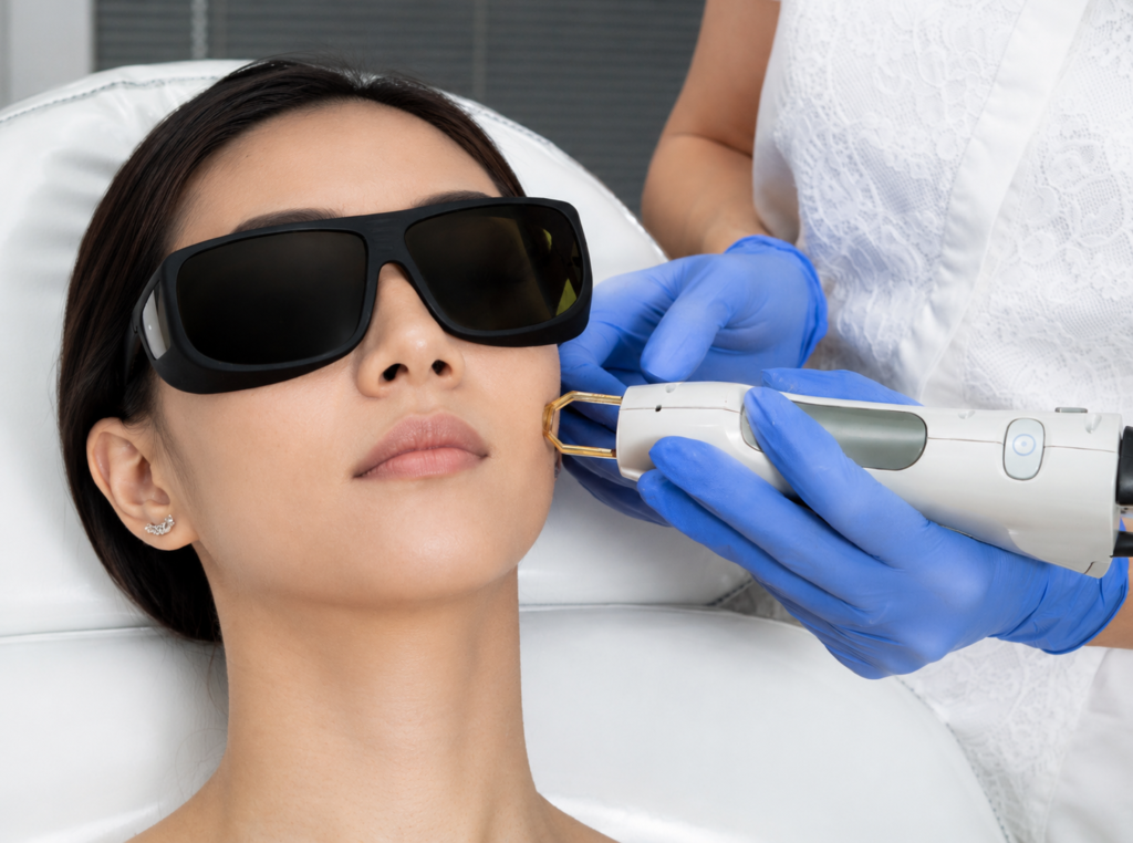
Pico laser has become the default for epidermal pigment in skin of colour because picosecond pulse durations fragment melanin with less heat and lower PIH risk than older nanosecond lasers. Useful for lentigines, freckles, and surface-layer melasma.
Q-switched Nd:YAG at 1064 nm remains the workhorse for dermal pigmentation, especially Hori’s nevus. It delivers energy deep enough to reach dermal melanocytes without harming the epidermis.
Fractional laser treats mixed pigmentation combined with texture issues, scarring, or dullness. Downtime is longer and PIH risk climbs, so settings need to be conservative for Singaporean skin.
A patient who asks for a pico laser on their “melasma” that is actually Hori’s nevus will pay for sessions that achieve less than they should. Matching laser to pigment is clinical judgement, not a menu choice.
Photoprotection is doing more work than patients realise
Tinted mineral sunscreens have emerged as the serious photoprotection choice for pigmentation management because iron oxides block visible light, which provokes melasma in a way traditional UV filters do not. A 2025 randomised investigator-blinded study published in the Journal of Cosmetic Dermatology compared a visible-light-protective tinted sunscreen against an untinted equivalent over five months in melasma patients with Fitzpatrick phototype III and IV. The tinted group showed better uniformity of pigmentation between affected and unaffected skin, with both products preventing sunlight-induced darkening when applied at least twice daily.
The practical takeaway is that a broad-spectrum SPF 50+ with iron oxide protection is the first product anyone serious about pigmentation should wear every day, and reapply every two to three hours outdoors. UVA passes through glass, which is why patients who sit beside office windows still develop pigmentation they thought they were shielded from.
What a realistic treatment plan looks like
Pigmentation treatment in Singapore usually runs in phases. A typical melasma protocol might start with eight weeks of topical hydroquinone plus oral tranexamic acid, move into gentle pico laser sessions spaced three to four weeks apart, then taper into maintenance with a long-term night-time brightening routine and strict daily photoprotection.
Solar lentigines often clear in one to three laser sessions. Hori’s nevus typically needs four to eight sessions spread across a year. PIH responds to topicals and time, with lasers used cautiously only after inflammation settles.
Costs vary by clinic and laser type. Public clinic pricing pages in 2025 show single pico laser sessions in Singapore ranging from around S$350 to S$800. Full melasma packages that bundle topicals, oral medication, and laser often sit between S$2,000 and S$5,000 across a complete treatment cycle.
Expectation-setting is where most treatment relationships succeed or fail. Clearance is not the same as cure. UV, hormones, and heat will keep doing what they do, and any honest plan includes that reality.
Conclusion
Pigmentation on Singaporean skin is rarely one condition in isolation. Melasma, sun damage, and PIH often stack together, shaped by a tropical UV load that never fully switches off. Accurate diagnosis matched to a sensible combination of topicals and lasers, backed by daily visible-light photoprotection, produces results that hold. Guesswork and aggressive DIY treatment usually make things worse.
Book a consultation with a qualified dermatologist or MOH-accredited aesthetic physician before starting any laser or prescription-strength topical. Then build a daily skincare regimen that supports the clinical work and protects the investment through Singapore’s climate. Explore the Skincode brightening range to anchor the topical side of your plan with formulations built for sensitive, reactive skin.
FAQ About Pigmentation Treatment Singapore
How long does pigmentation treatment in Singapore usually take to show results?
Epidermal pigmentation such as freckles and solar lentigines often responds within one to three pico laser sessions. Melasma and Hori’s nevus move more slowly, typically requiring three to six months of combined laser, topical, and photoprotection before clearance becomes visible.
Is melasma the same as Hori’s nevus?
No. Melasma is usually mixed epidermal and dermal pigmentation driven by hormones and UV exposure, while Hori’s nevus is purely dermal pigmentation caused by ectopic melanocytes in the dermis. A Wood’s lamp examination plus clinical history separates the two.
Can pigmentation return after laser treatment?
Yes. UV exposure, hormonal shifts, and heat reactivate melanogenesis, especially in melasma. Daily broad-spectrum SPF 50 with iron oxide visible-light protection, paired with a maintenance topical regimen, is the standard way to hold laser results.
Does hydroquinone work on every type of pigmentation?
Hydroquinone works best on epidermal pigmentation such as solar lentigines and surface-layer melasma. It has minimal effect on dermal pigmentation like Hori’s nevus, which requires laser intervention. Cycling is important to avoid ochronosis and rebound pigmentation.
Is pico laser safer than Q-switched laser for skin of colour?
Pico laser generally carries lower PIH risk than older Q-switched nanosecond lasers because of its shorter pulse duration and reduced thermal spread. Q-switched Nd:YAG at 1064 nm remains the correct choice for dermal pigmentation where deep penetration matters more than speed.
The information in this article is general and educational. It does not replace a personal consultation, diagnosis, or treatment from a qualified doctor. Please seek advice from a doctor about your individual skin condition.